Portal:Medicine/Selected picture archive
Archives
[edit]
Today, January 10, 2025, is in week number 2.
The following pictures were chosen for display on Portal:Medicine:
April 20, 2008 - April 27.
[edit]
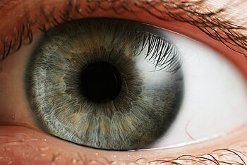
Photo credit: Llywrch
April 13, 2008 - April 20.
[edit]
Photo credit: Seldin MF, Shigeta R, Villoslada P, Selmi C, Tuomilehto J, et al. (2006) European Population Substructure: Clustering of Northern and Southern Populations. PLoS Genet 2(9): e143 Fig. 4(b)
April 6, 2008 - April 13.
[edit]
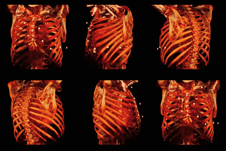
Photo credit: Original uploader was Che (CC-BY-2.5)
March 30, 2008 - April 6.
[edit]
Photo credit: Haymanj
March 23, 2008 - March 30.
[edit]
Photo credit: Someguy1221
March 16, 2008 - March 23.
[edit]
Photo credit: PD image from Gray's Anatomy, from bartleby.com.
March 9, 2008 - March 16.
[edit]
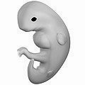
Photo credit: The present image (without watermark) was donated by Wouter Vergeer <wouter.vergeer@tribal.nl> on Wednesday, August 29, 2007 on behalf of 3DPregnancy.com.
March 1, 2008 - March 9.
[edit]
Photo credit: https://www.flickr.com/photos/euthman/304334264
February 25, 2008 - March 1.
[edit]
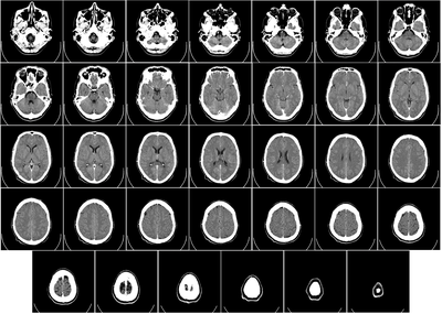
Photo credit: Radiology, Uppsala University Hospital. Brain supplied by Mikael Häggström. It was taken Mars 23, 2007, after an incidence of w:homonymous hemianopsia, but nothing strange was found. No further symptoms have appeared since then.
February 18, 2008 - February 25.
[edit]Photo credit: Public Domain
February 12, 2008 - February 18.
[edit]
Photo credit: Original uploader was User:Com4
February 4, 2008 - February 11.
[edit]
Photo credit: Original uploader was KieranMaher at en.wikibooks
January 28, 2008 - February 4.
[edit]
Photo credit: Original uploader was KieranMaher at en.wikibooks
January 21, 2008 - January 28.
[edit]
Photo credit: Original uploader was Jason7825 at en.Wikipedia
January 15, 2008 - January 22.
[edit]
Photo credit: Public Domain
January 8, 2008 - January 15.
[edit]
Photo credit: http://www.dodmedia.osd.mil/Assets/1991/Army/DA-ST-91-01841.JPEG
January 1, 2008 - January 7.
[edit]
Photo credit: Public Domain
