IL2RA
The interleukin-2 receptor alpha chain (also called Tac antigen, P55, and mainly CD25) is a protein involved in the assembly of the high-affinity interleukin-2 receptor, consisting of alpha (IL2RA), beta (IL2RB) and the common gamma chain (IL2RG). As the name indicates, this receptor interacts with interleukin-2, a pleiotropic cytokine which plays an important role in immune homeostasis.[5][6]
Genetics
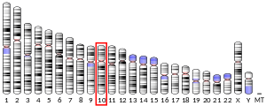
[edit]The human protein interleukin-2 receptor subunit alpha is encoded by a gene called IL2RA with a length around 51,6 kb. Alternative names for this protein coding gene are IL2R, IDDM10 and TCGFR. Location of IL2RA in human genome is on the short arm of 10th chromosome (10p15.1).[7][8][9]
Several frequent point mutations, single nucleotide polymorphism (SNP), have been identified in or in close proximity to IL2RA gene in the population. These SNPs have been linked mainly to susceptibility to immune dysregulation disorders, with majority found in research on multiple sclerosis (MS) and type 1 diabetes mellitus.[10][11][12][13][14]
IL2RA gene orthologues with identical protein functionality are relatively abundant and constant among animal species, especially in mammals subgroups. Moreover, conserved homologs of this gene are in mouse, rat, dog, cow, chimpanzee and Rhesus monkey.[15][16]
Expression
[edit]CD25 is expressed broadly among leukocytes. The highest surface expression of this protein is on regulatory T cells (Tregs), where CD25 is expressed constitutively, especially on a subset classified as naturally occurring Tregs. It can also be found on activated B cells, NK (natural killer) cells, thymocytes, and some myeloid lineage cells (e.g. macrophages, dendritic cells).[17][18] IL2RA has been used as a marker to identify CD4+FoxP3+ regulatory T cells in mice. However, there are species differences as CD25 is constitutively expressed by a large proportion of resting memory T cells non-regulatory CD4 T cells in humans that are absent in mice.[19][20] High expression of CD25 is also found on TCR activated conventional T cells (both CD8+ and CD4+ T lymphocytes), where it is considered to be a marker of T cell activation.[21] Additionally, expression of the IL-2 receptor alpha subunit can be found in non-lymphoid tissues such as lungs (alveolar macrophages), liver (Kupffer cells) and skin (Langerhans cells).[5][18]
IL2RA protein can be expressed in many types of neoplastic cells, such as in most B-cell neoplasms, T-cell lymphomas, some acute nonlymphocytic leukemias, neuroblastomas, mastocytosis, Waldenstrom macroglobuliaemia and tumor infiltrating lymphocytes.[22][23]
Structure
[edit]Interleukin-2 receptor alpha chain is an integral-membrane protein, more precisely type I transmembrane protein. This bitopic polypeptide is constructed by a sequence of 272 amino acids and has a molecular mass of around 30.8 kDa.[8] CD25 consists of three domains: extracellular (N-terminus), transmembrane (alpha-helix) and cytoplasmic (C-terminus). However, while extracellular part is able to function as a binding site for interleukin-2, short cytoplasmic domain lacks an ability to induce intracellular signalling and therefore needs to oligomerise with other IL-2 receptor subunits.[8][9] The interleukin-2 (IL2) receptor alpha (IL2RA) and beta (IL2RB) chains, together with the common gamma chain (IL2RG), constitute the high-affinity IL-2 receptor complex (Kd ~10−11M). Homodimeric alpha chains (IL2RA) result in low-affinity receptor (Kd ~10−8M) with no signalling ability, while dimeric beta (IL2RB) and gamma chains (IL2RG) produce a medium-affinity receptor (Kd ~10−9M). Moreover, CD25 is an exclusive subunit that entirely binds IL-2, while CD132 binds the shared γc family cytokines (IL-4, IL-7, IL-9, IL-15 and IL-21), and the CD122 subunit binds also IL-15.[5][24][25]
Soluble IL2RA has been isolated and determined to result from extracellular proteolysis during activation of T lymphocytes.[18] Also, alternately-spliced IL2RA mRNAs have been isolated, but the significance of each is currently unknown.[26]
Signalling cascade of interleukin-2 Receptor
[edit]Interleukin-2 can interact with intermediate-affinity dimeric IL-2 receptor, which consists of beta (CD122) and gamma (CD132) chains or with high-affinity trimeric complex, where also alpha subunit (CD25) constructs the IL-2 receptor and provides enhanced specific binding force. After activation of the receptor by its ligand, heterodimerisation of beta and gamma intracellular domains takes place.[5][24] This coupling of subunits brings together Janus kinases JAK1 and JAK3, considering their association with respective cytoplasmic parts of beta and gamma subunits. Downstream phosphorylation leads to initiation of three signalling pathways: JAK-STAT pathway, PI3K/Akt/mTOR pathway and Ras/Raf/MEK/ERK (MAPK) pathway. Regarding JAK-STAT pathway, particular signal transducers and activators of transcription participate in this signalling cascade: STAT5, STAT1 and STAT3 and after dimerisation, they translocate to nucleus to perform transcription factor functions. All three signalling pathways are important for diverse cellular regulations, in terms of increased survival (anti-apoptotic effect), proliferation and cell growth, transcriptional regulation and cell differentiation.[25][6] T lymphocytes are influenced by IL-2R signalling in case of CD4+ T helper subtype differentiation: promoting Th1, Th2, Th9, Tfr (T follicular regulatory cells) and suppressing Th17, Tfh ( T follicular helper cells). Additionally, strength of IL-2R signalling in CD8+ T cytotoxic lymphocytes may be connected to phenotypic fate of these cells for effector and memory T cells formation.[5][18][27]
Clinical significance
[edit]Roifman's group was the first to identify immunological consequences of CD25 loss and the patient has suffered from chronic infections and severe autoimmunity resembling Immune dysregulation, Polyendocrinopathy, Enteropathy, X-linked (IPEX) syndrome, caused by mutations in FOXP3 gene.[24]
CD25 as a biomarker
[edit]Levels of CD25 soluble form, called sIL-2Rα, has been connected to pathogenesis of autoimmune diseases and cancer. Since sCD25 is produced during immune activation, it is used as one of biomarkers to track disease progression and to indicate outcome for clinical disorders. Especially, it is a hallmark for hyper-activated immune system and cytokine storm, which may lead to multiple organ system failure.[28] In cancer, increased levels of this soluble protein are diagnostic marker for leukemia and lymphoma.[29] Furthermore, sIL-2Rα levels have some significance also in infectious diseases and transplantation. Higher serum levels were correlated with severity and need for hospitalisation of COVID-19 patients.[30] sIL-2Rα amount in plasma of HIV ( human immunodeficiency virus) positive patients has a correlation to HIV viral load and so to disease progression. Similarly in Chagas disease, caused by the protozoan Trypanosoma cruzi, patients have increased levels of sIL-2Rα and autoantibodies.[31] In regard to transplantation, higher levels of sCD25 may be used as a predictor of organ rejection and graft-versus-host disease (GVHD) for hematopoietic transplantations. Concerning CVD (cardiovascular diseases) soluble IL-2Rα has positive correlation with hypertension, type 2 diabetes mellitus, cardiac sarcoidosis, stroke and heart failure. For neurological disorders, high levels of sIL-2Rα are a sign for increased risk of developing schizophrenia.[18]
CD25 as a therapeutic target
[edit]Since Tregs express IL-2Rα subunit constitutively on the surface, some immunotherapeutic approaches try to use this information for selectivity.[28] NARA1 antibody is used in antitumour approaches to preferentially supplement interleukin-2 to conventional CD8+ T cells . NARA1 binds to the cytokine on the IL-2Rα binding site preventing binding to CD25. This complex should therefore interact with conventional T lymphocytes over T regulatory cells and thus increase cytotoxic activity without increasing suppressing activity in tumour environment.[32] Antibodies directly against CD25 have been altered to contain ‘activating’ Fc regions for the purpose of antibody-dependent cell-mediated cytotoxicity, in this case Treg depletion. Antibody marks a cell with IL-2Rα subunit on the surface, which is subsequently recognized and cleared by myeloid cell with Fc receptor.[5] Moreover, for treatment of multiple sclerosis, drug called daclizumab binds to IL2RA and so blocks high-affinity IL-2 receptors on recently activated T cells for interaction with IL-2 as well as IL-2 cross-presentation by dendritic cells.[33][34]
From the other side, treatment strategies for autoimmune and inflammatory diseases need selectivity for Tregs and suppression of immune system. IL-2Rα subunit expression on Tregs secures better sensitivity to IL-2. Therefore, administration of low doses of the cytokine preferentially stimulates T regulatory cells over others. Low-dose IL-2 therapy is used for graft-versus-host disease, type 1 diabetes mellitus, hepatitis C virus-induced vasculitis and systemic lupus.[5][6]
References
[edit]- ^ a b c GRCh38: Ensembl release 89: ENSG00000134460 – Ensembl, May 2017
- ^ a b c GRCm38: Ensembl release 89: ENSMUSG00000026770 – Ensembl, May 2017
- ^ "Human PubMed Reference:". National Center for Biotechnology Information, U.S. National Library of Medicine.
- ^ "Mouse PubMed Reference:". National Center for Biotechnology Information, U.S. National Library of Medicine.
- ^ a b c d e f g Spolski R, Li P, Leonard WJ (October 2018). "Biology and regulation of IL-2: from molecular mechanisms to human therapy". Nature Reviews. Immunology. 18 (10): 648–659. doi:10.1038/s41577-018-0046-y. PMID 30089912. S2CID 51939991.
- ^ a b c Zhou P (October 2022). "Emerging mechanisms and applications of low-dose IL-2 therapy in autoimmunity". Cytokine & Growth Factor Reviews. 67: 80–88. doi:10.1016/j.cytogfr.2022.06.003. PMID 35803833. S2CID 250200065.
- ^ Leonard WJ, Donlon TA, Lebo RV, Greene WC (June 1985). "Localization of the gene encoding the human interleukin-2 receptor on chromosome 10". Science. 228 (4707): 1547–1549. Bibcode:1985Sci...228.1547L. doi:10.1126/science.3925551. PMID 3925551.
- ^ a b c "IL2RA Gene - GeneCards | IL2RA Protein | IL2RA Antibody". www.genecards.org. Retrieved 2023-01-25.
- ^ a b "UniProt". www.uniprot.org. Retrieved 2023-01-25.
- ^ Buhelt S, Søndergaard HB, Oturai A, Ullum H, von Essen MR, Sellebjerg F (June 2019). "Relationship between Multiple Sclerosis-Associated IL2RA Risk Allele Variants and Circulating T Cell Phenotypes in Healthy Genotype-Selected Controls". Cells. 8 (6): 634. doi:10.3390/cells8060634. PMC 6628508. PMID 31242590.
- ^ Pourakbari R, Hosseini M, Aslani S, Ayoubi-joshaghani MH, Valizadeh H, Roshangar L, et al. (September 2020). "Association between interleukin 2 receptor A gene polymorphisms (rs2104286 and rs12722489) with susceptibility to multiple sclerosis in Iranian population". Meta Gene. 25: 100750. doi:10.1016/j.mgene.2020.100750.
- ^ Qu HQ, Montpetit A, Ge B, Hudson TJ, Polychronakos C (April 2007). "Toward further mapping of the association between the IL2RA locus and type 1 diabetes". Diabetes. 56 (4): 1174–1176. doi:10.2337/db06-1555. PMID 17395754.
- ^ Qu HQ, Bradfield JP, Bélisle A, Grant SF, Hakonarson H, Polychronakos C (December 2009). "The type I diabetes association of the IL2RA locus". Genes and Immunity. 10 (Suppl 1): S42 – S48. doi:10.1038/gene.2009.90. PMC 2805446. PMID 19956099.
- ^ Lowe CE, Cooper JD, Brusko T, Walker NM, Smyth DJ, Bailey R, et al. (September 2007). "Large-scale genetic fine mapping and genotype-phenotype associations implicate polymorphism in the IL2RA region in type 1 diabetes". Nature Genetics. 39 (9): 1074–1082. doi:10.1038/ng2102. PMID 17676041. S2CID 13940200.
- ^ "Gene: IL2RA (ENSG00000134460) - Orthologues - Homo_sapiens - Ensembl genome browser 108". www.ensembl.org. Retrieved 2023-01-25.
- ^ "IL2RA interleukin 2 receptor subunit alpha [Homo sapiens (human)] - Gene - NCBI". www.ncbi.nlm.nih.gov. Retrieved 2023-01-25.
- ^ Abel AM, Yang C, Thakar MS, Malarkannan S (2018-08-13). "Natural Killer Cells: Development, Maturation, and Clinical Utilization". Frontiers in Immunology. 9: 1869. doi:10.3389/fimmu.2018.01869. PMC 6099181. PMID 30150991.
- ^ a b c d e Li Y, Li X, Geng X, Zhao H (October 2022). "The IL-2A receptor pathway and its role in lymphocyte differentiation and function". Cytokine & Growth Factor Reviews. 67: 66–79. doi:10.1016/j.cytogfr.2022.06.004. PMID 35803834. S2CID 250252097.
- ^ Triplett TA, Curti BD, Bonafede PR, Miller WL, Walker EB, Weinberg AD (July 2012). "Defining a functionally distinct subset of human memory CD4+ T cells that are CD25POS and FOXP3NEG". European Journal of Immunology. 42 (7): 1893–1905. doi:10.1002/eji.201242444. PMID 22585674.
- ^ El-Maraghy N, Ghaly MS, Dessouki O, Nasef SI, Metwally L (August 2018). "CD4+CD25-Foxp3+ T cells as a marker of disease activity and organ damage in systemic lupus erythematosus patients". Archives of Medical Science. 14 (5): 1033–1040. doi:10.5114/aoms.2016.63597. PMC 6111364. PMID 30154885.
- ^ Shipkova M, Wieland E (September 2012). "Surface markers of lymphocyte activation and markers of cell proliferation". Clinica Chimica Acta; International Journal of Clinical Chemistry. 413 (17–18): 1338–1349. doi:10.1016/j.cca.2011.11.006. PMID 22120733.
- ^ Flynn MJ, Hartley JA (October 2017). "The emerging role of anti-CD25 directed therapies as both immune modulators and targeted agents in cancer". British Journal of Haematology. 179 (1): 20–35. doi:10.1111/bjh.14770. PMID 28556984.
- ^ Jones D, Ibrahim S, Patel K, Luthra R, Duvic M, Medeiros LJ (August 2004). "Degree of CD25 expression in T-cell lymphoma is dependent on tissue site: implications for targeted therapy". Clinical Cancer Research. 10 (16): 5587–5594. doi:10.1158/1078-0432.CCR-0721-03. PMID 15328201.
- ^ a b c Goudy K, Aydin D, Barzaghi F, Gambineri E, Vignoli M, Ciullini Mannurita S, et al. (March 2013). "Human IL2RA null mutation mediates immunodeficiency with lymphoproliferation and autoimmunity". Clinical Immunology. 146 (3): 248–261. doi:10.1016/j.clim.2013.01.004. PMC 3594590. PMID 23416241.
- ^ a b Peerlings D, Mimpen M, Damoiseaux J (2021). "The IL-2 - IL-2 receptor pathway: Key to understanding multiple sclerosis". Journal of Translational Autoimmunity. 4: 100123. doi:10.1016/j.jtauto.2021.100123. PMC 8716671. PMID 35005590.
- ^ "Entrez Gene: IL2RA interleukin 2 receptor, alpha".
- ^ Kalia V, Sarkar S (2018-12-20). "Regulation of Effector and Memory CD8 T Cell Differentiation by IL-2-A Balancing Act". Frontiers in Immunology. 9: 2987. doi:10.3389/fimmu.2018.02987. PMC 6306427. PMID 30619342.
- ^ a b Damoiseaux J (September 2020). "The IL-2 - IL-2 receptor pathway in health and disease: The role of the soluble IL-2 receptor". Clinical Immunology. 218: 108515. doi:10.1016/j.clim.2020.108515. PMID 32619646.
- ^ Janik JE, Morris JC, Pittaluga S, McDonald K, Raffeld M, Jaffe ES, et al. (November 2004). "Elevated serum-soluble interleukin-2 receptor levels in patients with anaplastic large cell lymphoma". Blood. 104 (10): 3355–3357. doi:10.1182/blood-2003-11-3922. PMID 15205267.
- ^ Kaya H, Kaji M, Usuda D (April 2021). "Soluble interleukin-2 receptor levels on admission associated with mortality in coronavirus disease 2019". International Journal of Infectious Diseases. 105: 522–524. doi:10.1016/j.ijid.2021.03.011. PMC 7942057. PMID 33711520.
- ^ Mengel J, Cardillo F, Pontes-de-Carvalho L (2016-05-13). "Chronic Chagas' Disease: Targeting the Interleukin-2 Axis and Regulatory T Cells in a Condition for Which There Is No Treatment". Frontiers in Microbiology. 7: 675. doi:10.3389/fmicb.2016.00675. PMC 4866556. PMID 27242702.
- ^ Sahin D, Arenas-Ramirez N, Rath M, Karakus U, Hümbelin M, van Gogh M, et al. (December 2020). "An IL-2-grafted antibody immunotherapy with potent efficacy against metastatic cancer". Nature Communications. 11 (1): 6440. Bibcode:2020NatCo..11.6440S. doi:10.1038/s41467-020-20220-1. PMC 7755894. PMID 33353953.
- ^ "Zinbryta Summary of Product Characteristics" (PDF). European Medicines Agency. 2016. Archived from the original (PDF) on 2018-06-14. Retrieved 2016-12-09.
- ^ Pfender N, Martin R (December 2014). "Daclizumab (anti-CD25) in multiple sclerosis" (PDF). Experimental Neurology. 262 (Pt A): 44–51. doi:10.1016/j.expneurol.2014.04.015. PMID 24768797. S2CID 32444028.
Further reading
[edit]- Kuziel WA, Greene WC (June 1990). "Interleukin-2 and the IL-2 receptor: new insights into structure and function". The Journal of Investigative Dermatology. 94 (6 Suppl): 27S – 32S. doi:10.1111/1523-1747.ep12875017. PMID 1693645.
- Waldmann TA (February 1991). "The interleukin-2 receptor". The Journal of Biological Chemistry. 266 (5): 2681–2684. doi:10.1016/S0021-9258(18)49895-X. PMID 1993646.
- Vincenti F (September 2004). "Interleukin-2 receptor antagonists and aggressive steroid minimization strategies for kidney transplant patients". Transplant International. 17 (8): 395–401. doi:10.1007/s00147-004-0750-3. PMID 15365604. S2CID 9244114.
External links
[edit]- Mouse CD Antigen Chart
- Human CD Antigen Chart
- CD4+FoxP3+ regulatory T cells gradually accumulate in gliomas during tumor growth and efficiently suppress antiglioma immune responses in vivo[dead link]
- Overview of all the structural information available in the PDB for UniProt: P01589 (Interleukin-2 receptor subunit alpha) at the PDBe-KB.


